
Pancreatic cancer is among the most aggressive malignancies. Owing to its subtle early symptoms and the lack of reliable diagnostic biomarkers, many patients are diagnosed at an advanced stage, underscoring the urgent need for improved precision diagnostic strategies. Aberrant protein glycosylation is closely associated with tumor development and progression and offers a promising avenue for identifying new cancer biomarkers. Polysialic acid (polySia), a biologically important glycan, is aberrantly expressed in various malignant tumors and has been linked to tumor progression and metastasis. However, existing glycan-recognition reagents, particularly monoclonal antibodies, face limitations in selectivity, affinity, and versatility, hampering the precise analysis of polySia and its development as a cancer biomarker.
Recently, Professor Zhen Liu’s research group at the School of Chemistry and Chemical Engineering, Nanjing University, in collaboration with Dr. Jian He’s team at Nanjing Drum Tower Hospital, developed a high-performance molecularly imprinted nanoparticle (nanoMIP) targeting polySia. Compared with the monoclonal antibody evaluated in this study, the material exhibited superior recognition selectivity and affinity, enabling efficient and precise polySia recognition in complex biological samples. Through reverse-microemulsion-confined surface imprinting and cladding, the researchers constructed a polySia ‘glycan superbinder’ with remarkable molecular recognition performance, including cross-reactivity below 10.1% and a dissociation constant (Kd) on the order of 10-8 M. The superbinder serves both as a fluorescent nanoprobe for imaging polySia in human tumor tissues and as an affinity capture reagent for enriching polysialylated proteins from human serum. Fluorescence imaging across multiple tumor types revealed prominent polySia signals in pancreatic cancer tissues. Subsequent colocalization analysis with CD56 highlighted polysialylated CD56 (polySia-CD56) as a potential diagnostic biomarker for pancreatic cancer.
Building on these findings, the team applied the nanoMIP to clinical serum analysis and established an affinity-enrichment and quantitative detection strategy for polysialylated proteins and polySia-CD56. Clinical sample analysis showed that polySia-CD56 offered stronger diagnostic performance than the conventional marker CA19-9 in the evaluated cohort, with complementary expression patterns between the two markers. Combining polySia-CD56 with CA19-9 achieved a diagnostic accuracy of 94.1% in the prediction dataset, highlighting the potential clinical value of this approach. Overall, the work not only introduces a glycan superbinder that outperforms the comparator monoclonal antibody in molecular recognition but also provides a strategy for using artificial recognition materials to uncover tumor-associated glycans and glycoform-specific biomarkers.
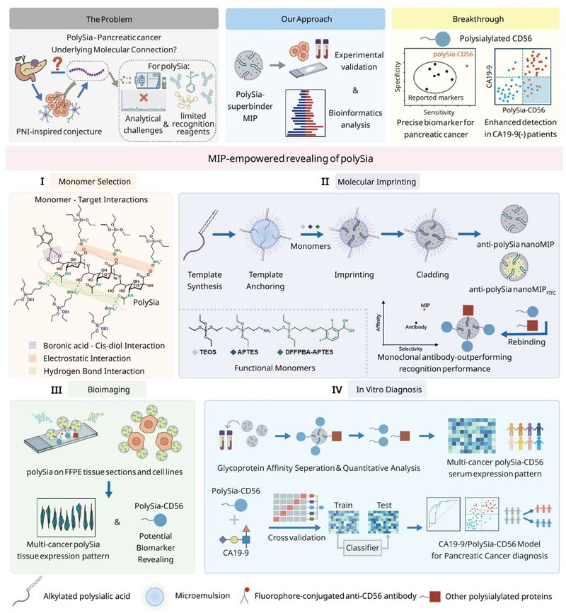
Figure 1. Schematic illustration of the construction of a polySia superbinder and its application in precision diagnosis of pancreatic cancer.
First, the researchers prepared anti-polySia molecularly imprinted nanoparticles (anti-polySia nanoMIPs) using reverse-microemulsion-confined surface imprinting and cladding. Rationally selected functional monomers enabled cooperative boronate affinity, electrostatic interactions, and hydrogen bonding for effective polySia recognition. The nanoparticles showed a relatively uniform size distribution and a clear imprinting effect. Selectivity and affinity measurements further demonstrated excellent polySia recognition, with cross-reactivity below 10.1% and a Kd on the order of 10-8 M. Both the affinity and selectivity surpassed those of the monoclonal antibody tested, underscoring the potential of the material as a high-performance glycan recognition tool (Figure 2).
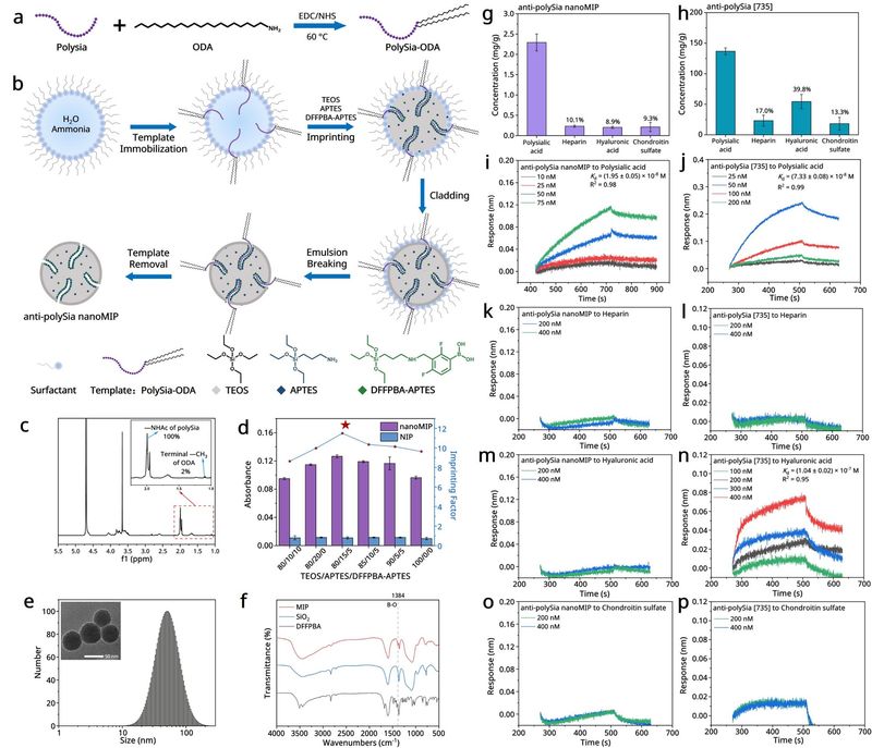
Figure 2. Preparation and characterization of the polySia superbinder and its recognition performance surpassing that of the comparator monoclonal antibody.
Next, using fluorescently labeled anti-polySia nanoMIPs, the researchers achieved specific fluorescence imaging of polySia in neuroblastoma cells and tumor tissues. They then extended the method to multiple types of human tumor tissues, combining histopathology with quantitative image analysis to investigate polySia expression patterns. Elevated signals were observed in neuroblastoma, brain glioma, pancreatic cancer, and certain lung and breast cancer tissues, whereas no significant differences were detected in liver, stomach, or colorectal cancer tissues. In particular, pancreatic tumor tissues displayed significantly stronger polySia fluorescence than adjacent non-tumor regions, suggesting an association between polySia and pancreatic cancer and providing a basis for subsequent biomarker discovery (Figure 3).
The team subsequently focused on pancreatic cancer tissues. Fluorescence imaging and quantitative analysis confirmed significantly increased polySia signals in tumor regions relative to adjacent non-tumor tissues, while competitive binding experiments further supported the specificity of nanoMIP recognition. Dual-channel fluorescence imaging, together with additional experimental validation, was used to investigate the molecular forms carrying polySia in pancreatic cancer tissues. Strong spatial colocalization between polySia and CD56, supported by other experimental evidence, indicated that polysialylated CD56 (polySia-CD56) represents an important form of polySia in these tissues, providing a foundation for developing a precision diagnostic biomarker (Figure 4).
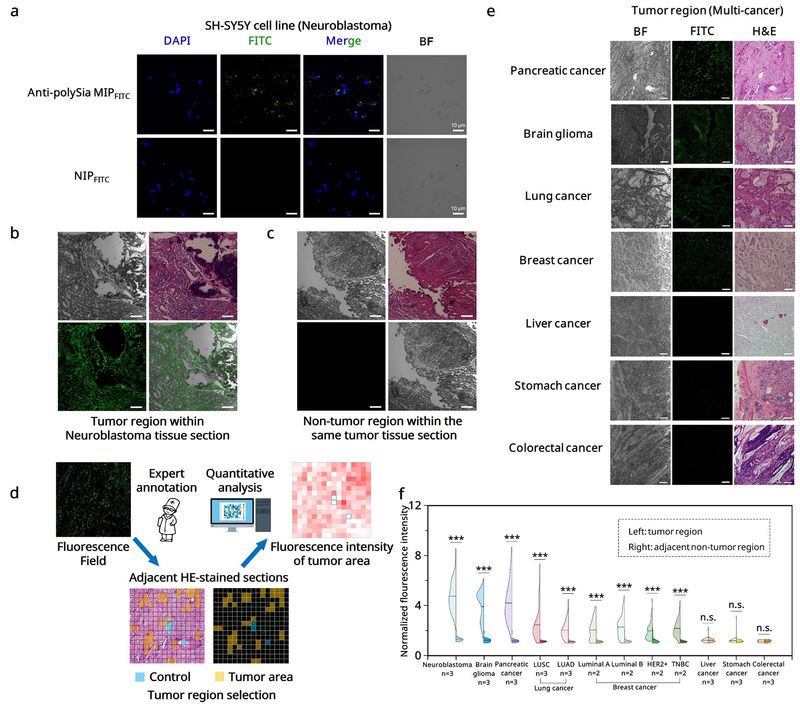
Figure 3. Fluorescence imaging and expression analysis of polySia in human tumor tissues using the glycan superbinder.
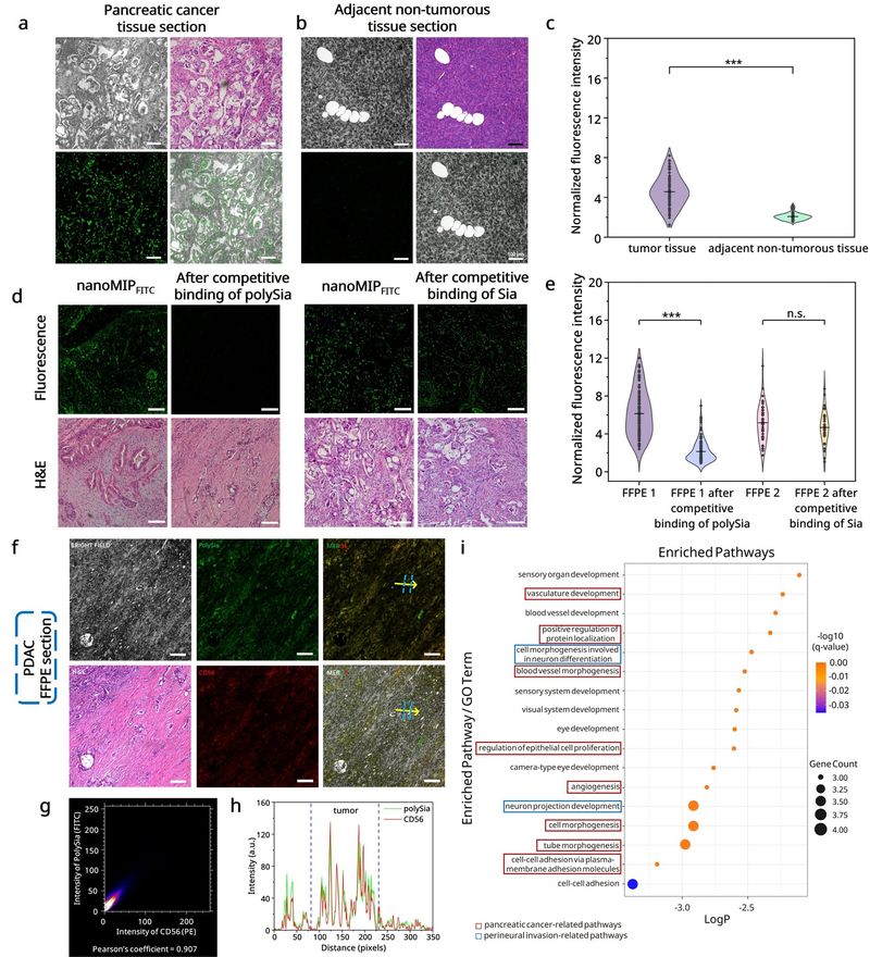
Figure 4. Preferential polySia expression in pancreatic cancer tissues and its colocalization with CD56.
Having established a potential association between polySia-CD56 and pancreatic cancer, the researchers exploited the excellent recognition properties of the nanoMIP to develop a serum assay based on affinity enrichment and quantitative analysis. Analysis of serum samples from patients with pancreatic cancer or other cancers, together with healthy controls, showed elevated polySia-CD56 levels in pancreatic cancer and a degree of discrimination among cancer types. Receiver operating characteristic (ROC) analysis further demonstrated promising performance in distinguishing pancreatic cancer patients from healthy individuals, supporting the potential of polySia-CD56 as a serum diagnostic biomarker (Figure 5).
The researchers then focused specifically on precision diagnosis of pancreatic cancer, systematically comparing polySia-CD56, total polysialylated proteins, total CD56, and the conventional marker CA19-9 in serum samples. PolySia-CD56 performed better than the other individual markers evaluated and showed a complementary expression pattern to CA19-9, helping identify some pancreatic cancer patients with negative CA19-9 results. Using machine learning, the team constructed a combined diagnostic model incorporating polySia-CD56 and CA19-9. The model achieved 94.1% diagnostic accuracy in the prediction dataset, further improving diagnostic performance. This result supports the potential of polySia-CD56 for precision diagnosis and offers a promising route to addressing limitations of CA19-9-based testing (Figure 6).
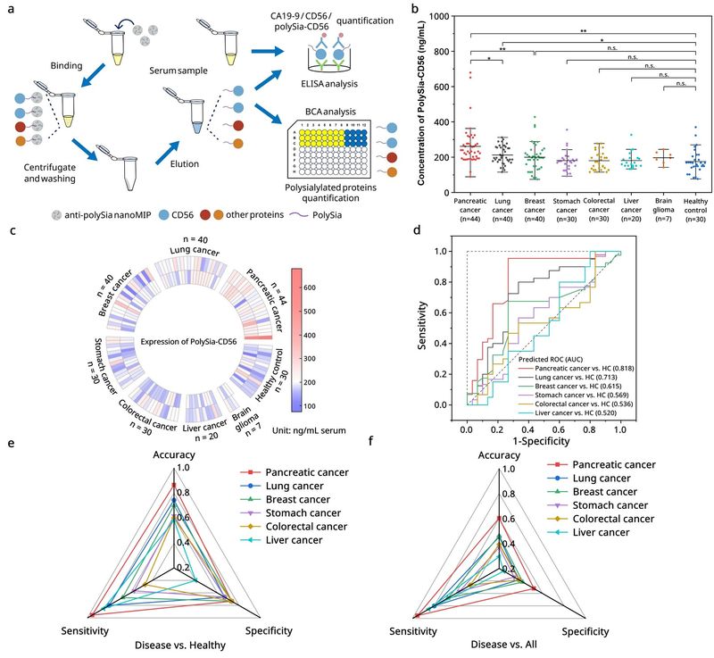
Figure 5. Serum detection of polySia-CD56 using molecular imprinting and evaluation of its specificity for pancreatic cancer diagnosis.
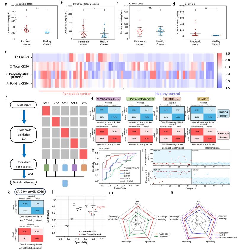
Figure 6. Complementary diagnostic advantages of polySia-CD56 and CA19-9 and performance of their combined assay.
This study, entitled ‘A Glycan Superbinder Reveals Polysialylated-CD56 as a Precision Biomarker for Pancreatic Cancer,’ is published in Proceedings of the National Academy of Sciences of the United States of America (PNAS), a leading multidisciplinary scientific journal. Professor Zhen Liu of the School of Chemistry and Chemical Engineering at Nanjing University and Dr. Jian He, Chief Physician in the Department of Medical Imaging and Nuclear Medicine at Nanjing Drum Tower Hospital, are the corresponding authors. This study was financially supported by the Key Grant (92478201) and National Major Scientific Instrumentation Development Project (22327804) from the National Natural Science Foundation of China and the Key Grant (5431ZZXM2404) from the State Key Laboratory of Analytical Chemistry for Life Science to Z.L.
Paper link:
https://doi.org/10.1073/pnas.2611747123